Pages

Showing posts with label Health. Show all posts

First robotic cervical surgery proves successful in US


A surgeon at the University of Texas Medical Branch at Galveston has performed a groundbreaking robotic laparoscopic procedure on a 35-year-old pregnant patient of Galveston, whose cervix was too short to sustain a pregnancy.
Dr. Sami Kilic, chief of minimally invasive gynecology and research at UTMB, is the first surgeon in the world reported to have used robotically assisted, ultrasound-guided laparoscopic surgery to successfully tighten a pregnant patient’s incompetent cervix.Kilic performed the surgery in December 2011 at UTMB’s John Sealy Hospital.
When performed traditionally, abdominal cerclage surgery requires a large incision and a long period of recovery.Kilic’s new procedure left the patient with only three tiny abdominal scars.
“The recovery was amazing. Two days later I was able to sit on the floor at home and play with my toddler,” new mum Leonora Orejuela said.Stitches to the cervix during surgery must be precise; a suture placed a hair’s breadth the wrong way can puncture either the amniotic sac or a major blood vessel in the mother.
With the dual visualization screen of the da Vinci Si robotic surgical system, Kilic was able to view a real-time ultrasound image on one screen and the operative field via scope camera on another screen, side by side, at the same time he performed the surgery.
This two-screen system offers unsurpassed visualisation in a laparoscopic surgical situation.
Kilic is an international pioneer in gynecologic robotic surgical techniques and training protocols who was hired by UTMB five years ago to spearhead a state-of-the-art robotic surgery training programme.
Orejuela, the patient, was discharged home the next day after a one-night stay in the hospital.
Orejuela proceeded to have an otherwise uncomplicated pregnancy, going into labor at 36 weeks.
She delivered a healthy baby girl, Lucia Munoz, 6 pounds, 11.5 ounces, by Caesarean section.
The procedure is published online in the Journal of Minimally Invasive Gynecology

Can your prenatal diet really help your baby's brain? Yes !! Read it!!!

Can your prenatal diet really help your baby's brain? More and more studies indicate YES! Research from the University of Connecticut (published in the June 2007 issue of American Journal of Clinical Nutrition) reported that infants of women who consumed DHA (a specific type of omega-3 fatty acid) during pregnancy performed better on problem-solving tasks. 
In the study, 29 pregnant women were randomly place into two groups at gestation week 24. One group received cereal bars containing 300 milligrams of DHA each. The second group of women received cereal bars containing no DHA. All study participants ate, on average, five bars each week for the remainder of their pregnancies. When their infants were nine months old, they were tested using the Infant Planning Test and the Fagan Test of Infant Intelligence. 
Lake trout


No significant differences in overall intelligence or memory recognition were noted between the two groups. However, the infants of the DHA-eating mothers performed significantly better in problem-solving.

According to these researchers, many pregnant women are not aware that consuming DHA is important during pregnancy and lactation. On top of that, women who are aware of their need for DHA are concerned with finding safe dietary sources of this omega-3 fatty acid. 
Fish and seafood, like salmon, lake trout, sardines, tuna, herring, oysters, shrimp and shellfish are major sources of DHA  for pregnant women .Nuts and seeds, such as walnuts, ground flaxseed, flaxseed oil Canola and soybean oils are also useful food to obtain DHA

wall nut

Docosahexaenoic acid (DHA) is an omega-3 fatty acid (molecular formula C22H32O2). In chemical structure, DHA is a carboxylic acid with a 22-carbon chain and six cis double bonds; the first double bond is located at the third carbon from the omega end. Its trivial name is cervonic acid, its systematic name is all-cis-docosa-4,7,10,13,16,19-hexa-enoic acid, and its shorthand name is 22:6(n-3) in the nomenclature of fatty acids.
DHA IUPAC name (4Z,7Z,10Z,13Z,16Z,19Z)-docosa-4,7,10,13,16,19-hexaenoic acid; Doconexent


DHA was also found to inhibit growth of human colon carcinoma cells. DHA concentrations in breast milk range from 0.07% to greater than 1.0% of total fatty acids, with a mean of about 0.34%. DHA levels in breast milk are higher if a mother's diet is high in fish. 
DHA is actively promoted by manufacturers as a food additive. Until recently, sales other than to makers of infant formula have been minimal; however, in 2007, several DHASCO-fortified dairy items (milk, yogurt) began selling in grocery stores.

Facts about Flesh-eating bacteria


Aimee Copeland, a Georgiagrad student, is fighting for her life because of the flesh-eating bacteria that infected her after she gashed her leg in a river two weeks ago. One of her legs was amputated and her fingers will be too, her father says, because of the spreading infection.
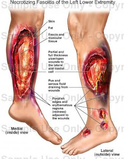
She has a rare condition, called necrotizing fasciitis, in which marauding bacteria run rampant through tissue. Affected areas sometimes have to be surgically removed to save the patient’s life.
Flesh Eating bacteria


HOW OFTEN DO PEOPLE GET THESE INFECTIONS?
The government estimates roughly 750 flesh-eating bacteria cases occur each year, usually caused by a type of strep germ.
However, Aimee Copeland’s infection was caused by another type of bacteria, Aeromonas hydrophila. Those cases are even rarer. One expert knew of only a few reported over the past few decades.
DO MOST PEOPLE SURVIVE?
Yes, but about 1 in 5 people with the most common kind of flesh-eating strep bacteria die. There are few statistics on Aeromonas-caused cases like Copeland’s.
HOW DOES SOMETHING LIKE THIS HAPPEN?
The germs that can cause flesh-eating disease are common in warm and brackish waters like ponds, lakes and streams. They are not a threat to most people. An infectious disease expert at Vanderbilt University, Dr. William Schaffner, said: “I could dive in that same stream, in the same place, and if I don’t injure myself I’m going to be perfectly fine. It’s not going to get on the surface of my skin and burrow in. It doesn’t do that.”
But a cut or gash — especially a deep one — opens the door for flesh-eating bacteria.
Foot seen after Infection

IS THERE ANYTHING YOU CAN DO TO AVOID SUCH AN INFECTION?
Prompt and thorough medical care should stop the infection before it spreads. A wound can look clean, but if it’s sutured or stapled up too soon it can create the kind of oxygen-deprived environment that helps these bacteria multiply and spread internally. Once established, these rare infections can be tricky to diagnose and treat.
Also, Aeromonas is resistant to some common antibiotics that work against strep and other infections, so it’s important that doctors use the best medicines.
ARE SOME PEOPLE MORE AT RISK?
Yes, people with weakened immune systems are. Copeland’s family has not said whether she had some type of medical condition that could have made her more vulnerable and relatives could not be reached for comment Monday. Her doctors, meanwhile, have refused interviews.
Copyright 2012 The Associated Press.

New Case of flesh-eating bacteria occurred in USA


After a case where student of psychology at the University of West Georgia was found infected by  the flesh eating bacteria known as Aeromonas hydrophila, A new case was seen in the USA and this time its New Mother.
   Lana Kuykendall is a paramedic, so when a rapidly spreading red and black bruise appeared on the back of her leg after giving birth to twins, she knew something was wrong.
She and husband, Darren, raced to the hospital and within 90 minutes, the new mother was having surgery for necrotizing fasciitis, a rare but potentially deadly infection also called the flesh-eating bacteria.
“She’s still critical,” Darren Kuykendall told GreenvilleOnline.com. “It’s been a nightmare.”
After a normal pregnancy, the Piedmont couple welcomed their twins, Abigail and Ian, on May 7 at an Atlanta hospital, he said. Except for Lana needing blood, the delivery went normally and the babies were fine, he said.
  Lana Kuykendall with Her Husband

But he said by the next day Lana, who had begun having leg cramps the night before, was weak. She couldn’t stand or walk. When tests revealed nothing wrong, they returned home Thursday.
By Friday morning, however, Lana, 36, discovered the strange lesion on the back of her left leg.
“That scared her. She thought it was a blood clot. So we rushed immediately to Greenville Memorial Hospital,” Darren Kuykendall said.
“And the longer she sat there, the bigger that spot got. It was initially the size of a 3-by-5 index card. But it got bigger and bigger. It moved a quarter of an inch in half an hour. Then the high-risk OB physician had a suspicion of what it was.”
Limbs Infeccted after Flesh eating bacteria attack it

Necrotizing fasciitis is a bacterial infection that often occurs in an arm or leg after a minor trauma or surgery, according to the National Necrotizing Fasciitis Foundation. Group A Strep is most often responsible in minor traumas, the group reports.
People have developed the condition through a host of experiences, including a C-section or natural childbirth, abdominal surgery or a scratch, a broken leg or a cut, according to the group.
A recent highly publicized case involves Aimee Copeland, a 24-year-old Georgia graduate student who got the infection after suffering a cut in a zip line accident, according to The Associated Press. Doctors had to amputate her left leg and may still have to remove her fingers, her father said.
The disease occurs when bacteria enter the body and emit toxins that destroy the soft tissue, which becomes infected and must be removed, according to the NF foundation. Along with surgery, treatment includes antibiotics and other medications.
If it spreads, it can cause systemic shock and death within days.
In Lana Kuykendall’s first surgery, doctors removed the dead skin and tissue, her husband said. A little more tissue was removed during a second and third surgery, but no more infected tissue was found in the last operation Monday, he said.
Group A strep caused the infection, which also spread to her blood, and she is being treated with antibiotics, he said. She was still on a ventilator in the intensive care unit on Tuesday and sedated.
Kuykendall, 42, has no idea how the infection occurred.
“They are saying things are leaning her way. Her vitals are good and her lab results are looking good,” he said. “But this could go either way at any given time.”
Married for four years, the couple met at the scene of an accident — he’s a firefighter and she’s a paramedic with Greenville Hospital System. The twins are healthy and being cared for by family and friends, he said.

College Girl Battles With Flesh Eating Bacteria!!!

A student of psychology at the University of West Georgia met with an accident when homemade zip line snapped and led her left calf open. Aimee Copeland, 24, received 22 stitches, but weeks after it was found that she has contracted the flesh eating bacteria known as Aeromonas hydrophila.
Aimee Copeland
She has lost her left leg and a part of abdomen and it is suspected that she will lose her hand fingers as well. Aimee's Father Andy Copeland said that Aimee is having remarkable recovery and instead of looking at the dark side of the story, they have been focusing on the brighter side.
Andy further affirmed that it feels great when they get to listen from doctors that Aimee is recovering well. Even doctors are shocked to see the determination of Aimee and they have affirmed that this determination is helping her to recover so well.
Aeromonas hydrophila  Image Taken from Electron Microscope


Dr. Buddy Creech, who is an Assistant Professor of pediatric infectious diseases at Vanderbilt University, said that the bacterium starts destroying the tissues that surround as it looks for nutrition. Destroying process leads to inflammation and swelling and once it comes, then it gets very difficult to control the infection.
"When it gets into those deeper tissues, it has a remarkable ability to destroy the tissues that surround it in sort of this hunt for nutrition", said Creech.
Affect seen After Bacteria slowly eat Muscles Tissue

New cavity-filling material reverses decay and regenerates tooth structure

A new composite material, which is made up of silver and calcium nanoparticles, could work as a dental filling that kills remaining bacteria so that patients don’t have to make a return trip to the dentist.
Dental fillings replace the part of the tooth drilled out inorder to remove decay. But if any bacteria remains, the cavity can grow right under the filling, Discovery News reported.
The new material, developed by researchers at theUniversity of Maryland, also rebuilds any structure affected by decay, essentially getting rid of the cavity altogether.

Due to their small size, the silver nanoparticles can invade the cellular structure of bacteria and other microorganisms and kill them. Calcium phosphate, also included in the composite, is responsible for building the tooth back up.
There have been questions raised about implementing these materials into toothpaste or mouthwash, but the scientific community isn’t ready to get on board with that just yet.
There is a lot of concern coming from scientists and researchers about the possible harmful affects of human consumption of the particles. Further testing will be conducted on volunteers to sort through the health concerns. 


Garlic can acts as stronger antibiotics for food-borne illness


Researchers have isolated a compound in garlic that is a 100 times more potent than popular antibiotics in combating Campylobacter bacteria, one of the commonest causes of intestinal illness. Some 2.4 million Americans alone are affected by Campylobacter every year, according to the Centers for Disease Control and Prevention, with symptoms including diarrhoea, cramping, abdominal pain and fever.
Garlic

"This work is very exciting to me because it shows that this compound (diallyl sulphide) has the potential to reduce disease-causing bacteria in the environment and in our food supply," says Xiaonan Lu, postdoctoral researcher at the Washington State University, who led the study.
Lu and colleagues looked at the ability of the garlic compound, diallyl sulphide, to kill the bacteria when it is protected by a slimy biofilm that makes it 1,000 times more resistant to antibiotics, the Journal of Antimicrobial Chemotherapy reports.
 They found the compound can easily penetrate the protective biofilm and kill bacterial cells by combining with a sulphur-containing enzyme, changing the enzyme's function and effectively shutting down cell metabolism, according to a Washington statement.
Diallyl Sulphide 


The researchers found that diallyl sulphide was as much as 100 times effective than much of the antibiotics erythromycin and ciprofloxacin and would often work in a fraction of the time.
"This is the first step in developing or thinking about new intervention strategies. Campylobacter is simply the most common bacterial cause of food-borne illness in the United States and probably the world," says Michael Konkel, study co-author who has been researching Campylobacter jejuni for 25 years.
Previously, Lu and colleagues found that diallyl sulphide effectively kills important foodborne pathogens, such as listeria monocytogenes and Escherichia coli O157:H7.

Believe it or not the British lab is growing human spare parts, now organ donation is a thing of the past


'This is a nose we’re growing for a patient next month,’ Professor Alexander Seifalian says matter-of-factly, plucking a Petri dish from the bench beside him.
Inside is an utterly lifelike appendage, swimming in red goo. Alongside it is another dish containing an ear. ‘It’s a world first,’ he says smiling.
‘Nobody has ever grown a nose before.’
His lab is little more than a series of worn wooden desktops strewn with beakers, solutions, taps, medical jars, tubing and paperwork, and looks like a school chemistry lab. 
But it’s from here that Seifalian leads University College London’s (UCL) Department of Nanotechnology and Regenerative Medicine, which he jokingly calls the ‘human body parts store’. 
Seifalian showing Nose made from nanomolecules

As he takes me on a tour of his lab I’m bombarded with one medical breakthrough after another. Daily Mail Reporter Said
At one desk he picks up a glass mould that shaped the trachea – windpipe – used in the world’s first synthetic organ transplant. 
At another are the ingredients for the revolutionary nanomaterial at the heart of his creations, and just beyond that is a large machine with a pale, gossamer-thin cable inside that’s pulsing with what looks like a heartbeat. It’s an artery. 
‘We are the first in the world working on this,’ Seifalian says casually to daily mail reporter. ‘We can make a metre every 20 seconds if we need to.’
‘Other groups have tried to tackle nose replacement with implants but we’ve found they don’t last,’ says Adelola Oseni, one of Seifalian’s team.
‘They migrate, the shape of the nose changes. But our one will hold itself completely, as it’s an entire nose shape made out of polymer.’
Looking like very thin Latex rubber, the polymer is made up of billions of molecules, each measuring just over one nanometre (a billionth of a metre), or 40,000 times smaller than the width of a human hair. Working at molecular level allows the material itself to be intricately detailed. 
Ear made in lab 

‘Inside this nanomaterial are thousands of small holes,’ says Seifalian.
‘Tissue grows into these and becomes part of it. It becomes the same as a nose and will even feel like one.’
When the nose is transferred to the patient, it doesn’t go directly onto the face but will be placed inside a balloon inserted beneath the skin on their arm. 
After four weeks, during which time skin and blood vessels can grow, the nose can be monitored, then it can be transplanted to the face.
At the cutting edge of modern medicine, Seifalian and his team are focusing on growing replacement organs and body parts to order using a patient’s own cells. There would be no more waiting for donors or complex reconstruction – just a quick swap. 
And because the organ is made from the patient’s own cells, the risk of rejection should, in theory, be eliminated.
Unsurprisingly, the recipe for the breakthrough biocompatible material used is a closely guarded secret. 
From those who have lost noses to cancer to others mutilated by injury, it’s hoped this revolutionary process could transform thousands of lives. 
‘We seed the patient’s own cells on to the polymer inside a bioreactor,’ says Oseni. 
This is a sterile environment mirroring the human body’s temperature, blood and oxygen supply. 
‘As the cells take hold and multiply, so the polymer becomes coated. The same methods could be applied to all parts of the face to reconstruct those of people who have had severe facial traumas.’
 ‘The full success of these implants needs to be tested with a larger number of patients in clinical trials,’ says Seifalian.
Such is the speed of progress that regenerative medicine is now moving on from replacing heart valves and rebuilding faces to potentially curing blindness and accelerating the study of some of the most debilitating diseases. 
The UK is at the forefront of this research, with work on a £54 million MRC Centre for Regenerative Medicine in Edinburgh completed earlier this year.
Until recently, regenerative medicine focused mostly on embryonic stem cells as these were the most versatile. They are called pluripotent, meaning they have the ability to become any cell type – blood, muscle, etc. 
By contrast, adult stem cells can replicate themselves endlessly, but only as the cell they began life as – skin cells replicate as skin cells, muscle cells as muscle cells.
But the moral debate surrounding embryonic stem cell research is controversial. 
Stem cells are taken from human embryos, which are destroyed in the process. 
In 2007, Professor Shinya Yamanaka of Kyoto University managed to create pluripotent cells from adult stem cells, potentially removing the need for embryonic stem cells completely.
These are known as induced pluripotent stem cells, or iPSCs. He was in part inspired by Professor Ian Wilmut, who was knighted for his role in the creation of Dolly the cloned sheep.
‘In the same way Dolly made us think maybe we could change cells, Yamanaka proved it could be done,’ says Wilmut.
‘This makes you think you can produce any cell type, producing nerves or muscle from skin cells, for example.’
This has been proved recently with the news that scientists at Cambridge Universityhave created brain cells from skin cells which could help with the search for new treatments for Alzheimer’s, stroke and epilepsy.
Sitting on a desk inside Seifalian’s laboratory is the mould for the trachea which he and his team created. It was recently implanted into a patient making it the world’s first ever synthetic organ transplant. 
The patient in question, a 36-year-old Eritrean man, had a large cancerous tumour in his throat that was rapidly spreading towards his lungs. The transplant was successful, and the patient is now out of hospital and recovering well.
On another bench in the lab lies an ear ready for seeding, while next door the team is working on heart valves that won’t even need seeding before implantation, having been developed instead to attract the cells they need once implanted. 
This will allow them to grow in the body instead of bioreactors and, along with an insertion method that removes the need to open the chest, could revolutionise heart bypass surgery.
‘Normally for heart bypass you take a section of vein from the patient’s leg or arm. But 30 per cent of patients don’t have suitable veins so can’t have the operation. No alternative currently exists for them,’ says Seifalian. 
‘We are the first in the world with this. Nobody else is even close. It has been successful in animal trials; this year it will be going for patient trials’.
While Seifalian and his team keep developing potential implants, on the other side of Londonanother team led by Professor Pete Coffey, the London Project to Cure Blindness, is using stem cells to tackle age-related macular degeneration, the most common form of age-related sight loss, which affects 513,000 people in the UKalone. 
‘There’s nothing that can be done for those with the disease,’ says Coffey. ‘There’s a real unmet need here.’
The aim is to replace the diseased cells with healthy new ones, restoring vision. 
Unlike Seifalian’s team, Coffey’s is using embryonic stem cells because in every experiment to date they are the only ones that work.
On the issue of working with embryonic stem cells Coffey is clear.
‘One thing I always face is that the term embryo has a different meaning for different people.
'The embryo in this case is five days old, and I know under various religious definitions that’s life, but I see this as similar to organ donation. That embryo cannot survive on its own.’
Most embryonic cells used in research, including Coffey’s, are from IVF treatment where a large surplus of embryos is part of the process. Unwanted embryos can be donated to research, otherwise, as Coffey says, ‘they’re disposed of.’
 ‘A human embryonic cell keeps reproducing itself naturally, so one cell generates everything we need – we’ve banked the duplicates in nitrogen chambers in three different countries – which means this cell could service a clinical population of 28 million. Isn’t that worth it?’
Coffey’s project is perhaps the most advanced major regenerative medicine project in the world today, scheduled for clinical trials with patients later this year. But even success in a patient trial is no guarantee a treatment will ever reach the mass market.
‘The sad thing is the time frame here,’ says Paul Whiting, executive director of Pfizer’s regenerative medicine arm, who is working closely with Coffey’s project. 
‘Even things that seem close are probably ten years away, while many are 20 to 50 years away. We need to know if these things will do long-term harm before they can reach patients, so it will be a gradual progression over at least 50 years.’
And a recent study illustrates just how far the divide between laboratory success and clinical reality could be: researchers at California University have found that mice treated with iPSCs made from their own skin cells ultimately reject the transplants.
When asked about this, Wilmut agrees it was valid but also says it was ‘a very preliminary observation’, another piece of the puzzle leading toward full understanding of the subject. There are also concerns that the reprogramming process used to create iPSCs might cause cancer in those same cells.
But back in Seifalian’s labs, the raw energy remains. 
‘Before, the idea was you rob Peter to pay Paul, taking one bit of the body to reconstruct another, but now the idea of being able to grow tissues in a lab and to reconstruct the body is huge,’  says Adelola Oseni.
‘If we can grow a heart, a lung or a trachea in a lab, we don’t need to wait for donors. 
'This work has massive implications for the way we function as clinicians and the way medicine is practised.’
Source : Daily Mail 


New method can make even muddy water safe for drinking


A scientist at Michigan Technological Universityhas developed a simple, cheap way to make water safe to drink, even if it's muddy. It's easy enough to purify clear water. The solar water disinfection method, or SODIS, calls for leaving a transparent plastic bottle of clear water out in the sun for six hours.
That allows heat and ultraviolet radiation to wipe out most pathogens that cause diarrhea, a malady that kills 4,000 children a day in Africa.
It's a different story if the water is murky, as it often is where people must fetch water from rivers, streams and boreholes.

 'In the developing world, many people don't have access to clear water, and it's very hard to get rid of the suspended clay particles,' said Joshua Pearce, an associate professor of materials science and engineering.
'But if you don't, SODIS doesn't work. The microorganisms hide under the clay and avoid the UV,' he explained.
Thus, to purify your water, you first have to get the clay to settle out, a process called flocculation.
Working with student Brittney Dawney of Queen's University in Ontario, Pearce discovered that one of the most abundant minerals on Earth does this job very well: sodium chloride, or simple table salt.
 Salt is inexpensive and available almost everywhere. And it doesn't take very much to make muddy water clear again.
 'The water has a lower sodium concentration than Gatorade,' Pearce said.
 This would still be too much salt to pass muster as American tap water, but American tap water is not the alternative.
 'I've drunk this water myself. If I were somewhere with no clean water and had kids with diarrhea, and this could save their lives, I'd use this, no question,' he added.
Salt works best when the suspended particles are a type of clay called bentonite. The technique doesn't work as well with other kinds of clay.
However, by adding a little bentonite with the salt to water containing these different clays, most of the particles glom together and settle out, creating water clear enough for SODIS treatment.
Pearce and Dawney are running more tests on water containing various types of clays, and they are also investigating different soil types across Africa to see where their methods might work the best.
Their paper has been accepted for publication in the Journal of Water, Sanitation, and Hygiene for Development and will appear in June. 

Daily glass of pomegranate boosts sexual desire in Both man and Women


Do you use Viagra as you don’t have sexual desire if Yes leave the Viagra and start drinking pomegranate Juice Every day it doesn’t only increases Sexual desire but also save us Heart disease and Cancer
Men and women who drank a daily glass of the fruit’s juice for a fortnight experienced a surge in the hormone testosterone, which increases sexual desire in both genders
The latest achievement claimed for pomegranates, which are already acclaimed as a superfood, will be welcome among those who would prefer a natural aphrodisiac.
The study, by researchers from Queen MargaretUniversity in Edinburgh, involved 58 volunteers aged between 21 and 64.
By the end of the fortnight both sexes had seen ‘significantly increased’ testosterone levels.
For men this affects traits such as facial hair, a deep voice and greater sexual urges.
It is also produced in female adrenal glands and ovaries, raising a woman’s sex drive and strengthening bones and muscles.
As a side effect, increased testosterone can help raise mood and memory and even relieve stress such as ‘pre-match nerves’ or stage fright, said the study.
Previous research on pomegranate juice has found it full of antioxidants which can help ward off heart diseases and help blood circulation.
The superfood’s ingredients also help fight various forms of cancer, alleviate the symptoms of osteoarthritis and cure stomach upsets and even conjunctivitis.
The Edinburgh research measured testosterone levels, blood pressure and, using a scientific scale, levels of 11 emotions including fear, sadness, guilt, shyness and self-assurance.
Testosterone levels increased between 16 per cent and 30 per cent among the subjects, while blood pressure plummeted.
Positive emotions rose and negative feelings fell.

Blind man can see the world after implanting Bionic Eye Microchips


'I've dreamed in colour for the first time in 20 years': Blind British man can see again after first successful implant of 'bionic' eye microchips
It was the ‘magic moment’ that released Chris James from ten years of blindness.
Doctors switched on a microchip that had been inserted into the back of his eye three weeks earlier.
After a decade of darkness, there was a sudden explosion of bright light – like a flash bulb going off, he says.
Now he is able to make out shapes and light. He hopes his sight – and the way his brain interprets what the microchip is showing it – will carry on improving.
Mr James, 54, is one of two British men who have had their vision partly restored by a pioneering retina implant.
Chip of 3mm by 3mm that is planted to sufferers Eye
The other, Robin Millar, one of Britain’s most successful music producers, says he has dreamed in colour for the first time. 

Both had lost their vision because of a condition known as retinitis pigmentosa, where the photoreceptor cells at the back of the eye gradually cease to work.
Their stories bring hope to the 20,000 Britons with RP – and to those with other eye conditions such as advanced macular degeneration which affects up to half a million.
Mr James had a ten-hour operation to insert the wafer-thin microchip in the back of his left eye at the Oxford University Eye Hospital six weeks ago. Three weeks later, it was turned on..
Mr James, who lives in Wroughton, Wiltshire, with his wife Janet, said of his ‘magic moment’: ‘I did not know what to expect but I got a flash in the eye, it was like someone taking a photo with a flashbulb and I knew my optic nerve was still working.’
Chip Pairs with This External Device to Process Image

The microchip has 1,500 light sensitive pixels which take over the function of the retina’s photoreceptor rods and cones.
One of the first tests was making out a white plate and cup on a black background.
 Mr James, who works for Swindon Council, said to Daily mail ‘It took a while for my brain to adjust to what was in front of me, but I was able to detect the curves and outline of these objects.’
Tim Jackson, a consultant retinal surgeon at King’s College Hospital and Robert MacLaren, a professor of ophthalmology at the University of Oxford and a consultant retinal surgeon at the Oxford Eye Hospital, who are running the trial, say it has ‘exceeded expectations’ with patients already regaining ‘useful vision’.

  Ten more Britons with RP will be fitted with the implants, which are also being tested in Germanyand China. The device, made by Retina Implant AG of Germany, connects to a wireless power supply buried behind the ear. This is connected to an external battery unit via a magnetic disc on the scalp. The user can alter the sensitivity of the device using switches on the unit.
Mr Jackson said: ‘It’s difficult to say how much benefit each patient will get, this pioneering treatment is at an early stage.
Surgeon Robert Mclaren

‘But it’s an exciting and important step forward. Many of those who receive this treatment have lost their vision for many years. The impact of them seeing again, even if it is not normal vision, can be profound and at times quite moving.’ Mr Millar, 60, who was behind Sade’s Diamond Life album, has been blind for 25 years. He said: ‘Since switching on the device I am able to detect light and distinguish the outlines of objects.
‘I have even dreamt in very vivid colour for the first time in 25 years so a part of my brain which had gone to sleep has woken up! I feel this is incredibly promising and I’m happy to be contributing to this legacy.’

Baldness can be cured using stem cells


Are you bald and having trouble facing people? If yes your worries are over as Japanese researchers have successfully grown hair on hairless mice by implanting follicles created from stem cells, they announced Wednesday, sparking new hopes of a cure for baldness.
Led by Professor Takashi Tsuji from Tokyo University of Science, the team bioengineered hair follicles and transplanted them into the skin of hairless mice.
The creatures eventually grew hair, which continued regenerating in normal growth cycles after old hairs fell out.
When stem cells are grown into tissues or organs, they usually need to be extracted from embryos, but Tsuji and his researchers found hair follicles can be grown with adult stem cells, the study said.
Baldness can now be cured

“Our current study thus demonstrates the potential for not only hair regeneration therapy but also the realization of bioengineered organ replacement using adult somatic stem cells,” it said.
The combination of the new and existing technologies is expected to improve treatment for baldness, possibly allowing people to use their own cells for implants that will give them their hair back.
“We would like to start clinical research within three to five years, so that an actual treatment to general patients can start within a decade,” said researcher Koh-ei Toyoshima.

Doctors remove four extra limbs from baby who was born with


A baby boy born with six legs has had a successful operation to remove his four extra limbs, doctors said today.
The youngster from Karachi in Pakistanwas believed to have had a parasitic twin, which had not developed properly in the womb, resulting in the extra legs.
A team of five doctors had fought to save the boy's life at the National Institute of Child Health in Karachi.
Baby Boy after Surgery
The head of the NICH, Jamal Raza, said the abnormal birth was the result of a genetic disease which would affect only one in a million or more babies.
'It was strange that apparently an abnormal baby with six legs was as normal as other children,' he said.
Before surgeons could operate they said they had to work out which of the limbs belonged to the boy and which to his twin.
Baby Before surgery 

Doctors examined MRI, blood tests and CT scan reports before deciding to perform the surgery. The operation lasted eight-hours and was performed in stages.
The baby had been in an intensive care unit ward since he was born last week to the wife of an X-ray technician.

Imran Shaikh, the baby's father who lives in Sukkur, said he was grateful his son was treated.
'We are a poor family. I am thankful to the government and doctors for helping us successful operating my baby,' he said.
Shaikh and his wife live in Sukkur, around 280 miles north of where his son is being cared for. His wife is reported to be recovering well from the birth and in a good state of health. 
His wife - who is also his cousin - has been unable to travel because she had a caesarean section delivery.
He said they were planning on naming their son Umar Farooq.
The Sindh provincial health department said they were examining if he needed any further treatment to live a normal life.

Doctors battle to save baby born with SIX legs


Pakistani doctors are battling the odds to save a newborn baby born with a rare genetic condition that has left him with six legs. 
The one-week-old boy is believed to be one of parasitic twins. 
His conjoined twin was born prematurely and incompletely developed, which resulted in the second child having the extra legs, said Jamal Raza, director of the National Institute of Child Health in Karachi, to News.com.
Doctors at the institute are fighting to save the newborn, who remains in an intensive care unit ward. 
Baby born with six legs 

Raza added that they were planning to operate on the boy and were considering asking for help from foreign experts with more experience in the rare disease, believed to afflict just one in one million babies. 
He tried to clarify that the baby did not have six legs – he had two legs and the other four belonged to the other twin.
'Operating on such a baby is not an easy task as proper assessments need to be done first,' he said. 'We need to figure out whether the baby has his twin’s limbs or his own. We also need to consider how much the internal organs have developed as the latter could complicate matters and decrease the baby’s chances of surviving.'
Baby boy born with six legs to be names Umar Farooq

Speaking to the news site, the baby's father, Imran Shaikh, made a plea for help from the government and charities.
‘I can’t afford to visit Karachi and get treatment for my baby,’ said the X-ray technician, who earns the equivalent of $66-a-month. ‘I appeal to philanthropists and the government to come forward for the treatment.’
Shaikh and his wife of four years live in Sukkur, around 280 miles north of where his son is being cared for. His wife is reported to be recovering well from the birth and in a good state of health. 
His wife - who is also his cousin - has been unable to travel because she had a caesarean section delivery.

He said they were planning on naming their son Umar Farooq.
Muhammad Qaisar, a doctor at Pakistan Institute of Medical Sciences in Islamabad, told allvoices.com he was certain the boy could be successfully operated on. ‘It is perhaps [the] first child in the history of Pakistan having six legs,’ he said.
‘The case will also be a test for doctors and we hope for the better,” he said.
Since Shaikh made his public plea for help, the Sindh Governor has come forward and directed officials concerned to make sure the child receives all the medical care he needs, Pakistani-based The Nation reported. 

Treatment of HIV aids possible now, engineered stem cells kill HIV


UCLA researchers say they've shown that genetically engineered stem cells can attack HIV-infected cells in a living organism. The 'warrior' cells have been shown to seek out and destroy HIV in mice.
"We believe that this study lays the groundwork for the potential use of this type of an approach in combating HIV infection in infected individuals, in hopes of eradicating the virus from the body," says UCLA assistant professor of medicine Scott Kitchen.


 The scientists had already identified the molecule known as the T cell receptor - which guides the T cell in recognizing and killing HIV-infected cells - cloned it and used it to genetically engineer human blood stem cells.
When these cells were placed in human thymus tissue that had been implanted in mice, they blossomed into a large population that could specifically target cells containing HIV proteins.
Now, the researchers have engineered human blood stem cells in much the same way and found that they can form mature T cells that can attack HIV in tissues where the virus resides and replicates.
Drugs used now to control the damage by Virus
In a series of tests on the mice's peripheral blood, plasma and organs, they found that the number of CD4 'helper' T cells — which die off as a result of HIV infection — increased, while levels of HIV in the blood decreased.
This shows that the engineered cells were able to develop and migrate to the organs to fight infection there.

"We believe that this is the first step in developing a more aggressive approach in correcting the defects in the human T cell responses that allow HIV to persist in infected people," says Kitchen.

The researchers will now begin making T cell receptors that target different parts of HIV and that could be used in more genetically matched individuals, he says

Share

Twitter Delicious Facebook Digg Stumbleupon Favorites More